Herbal Remedies of Hearing loss in children – Causes, Symptoms, Diagnosis & Treatment
Abstract
Pediatric hearing loss presents a critical developmental challenge, directly compromising speech acquisition, cognitive maturation, psychosocial integration, and scholastic achievement. From a modern clinical perspective, pediatric hearing impairment is broadly categorised into conductive, sensorineural, or mixed hearing loss, stemming from genetic, congenital, infectious, or environmental factors. WHO emphasises prevention, early identification, and rehabilitation as key components of childhood hearing care. In classical Ayurvedic literature, hearing loss is described as Badhirya or Karnabadhirya, classified under the twenty-eight Karna Rogas (diseases of the ear) by Acharya Sushruta and identified as one of the eighty Nanatmaja Vata Vyadhis (diseases caused exclusively by Vata dosha) by Acharya Charak. This article provides a holistic synthesis of pediatric hearing loss, exploring its aetiology, pathophysiology (Samprapti), clinical manifestations, and conventional diagnostic methodologies alongside traditional Ayurvedic concepts.
Introduction
Hearing is one of the fundamental sensory gateways through which a child interacts with, interprets, and learns from their environment. Hearing impairment during early childhood—particularly during the critical language-acquisition window from birth to five years—can lead to profound delays in verbal communication, emotional bonding, and intellectual growth. Global epidemiological data indicate that one to three per thousand live births present with congenital hearing loss, while many more develop acquired or progressive auditory deficits during early childhood. While modern medicine relies predominantly on surgical interventions such as tympanostomies, ossiculoplasties, and cochlear implants, or mechanical amplification via hearing aids, complementary holistic approaches are increasingly sought to optimise auditory nerve function, manage middle ear pathologies, and enhance overall quality of life. Ayurveda, the traditional science of medicine, offers a nuanced framework for understanding auditory dysfunction. Viewing the ear (Shrotrendriya) as a principal seat of Vata dosha located within the head (Uttamanga), classical Ayurvedic texts provide a thorough understanding of aetiology, structural impairment, and channel obstruction, leading to structured, individual-centric therapeutic protocols.
What Is Childhood Hearing Loss?
Hearing loss occurs when the auditory system cannot detect or process sound normally. The auditory system includes the external ear, middle ear, cochlea, auditory nerve, and central auditory pathways. It is generally classified into three major categories.
- Conductive hearing loss occurs when sound cannot travel efficiently through the external or middle ear. Earwax obstruction, middle-ear fluid, ear infections, perforation of the eardrum, or structural abnormalities can contribute to this form.
- Sensorineural hearing loss develops when the cochlea or auditory nerve is affected. It may be congenital or acquired and can be permanent.
- Mixed hearing loss contains both conductive and sensorineural components.
Another clinically important distinction is between unilateral and bilateral hearing loss. Even hearing impairment affecting only one ear may make it more difficult for a child to locate sounds or understand speech in noisy environments.
Causes of Hearing Loss in Children
Childhood hearing impairment has multiple possible causes, and identifying the underlying cause helps determine management.
Genetic Factors
Genetic conditions are an important cause of congenital or early-onset sensorineural hearing loss. Hearing impairment may occur independently or as part of a syndrome involving other organs. A child may have no obvious external abnormality despite having an inherited alteration affecting cochlear development or function.
Congenital Infections
Certain infections acquired during pregnancy can affect the developing auditory system. Congenital cytomegalovirus is an important example. Other infections, including rubella, can also contribute to childhood hearing impairment. Vaccination and appropriate maternal healthcare can reduce the risk associated with several preventable infectious causes.
Prematurity and Birth-Related Complications
Premature birth, very low birth weight, severe neonatal jaundice, prolonged intensive care, and certain complications around delivery may increase the risk of hearing impairment. These children may therefore require closer developmental and hearing surveillance.
Middle-Ear Disease
Otitis media and persistent fluid behind the eardrum are common causes of conductive hearing loss in childhood. The hearing reduction may fluctuate and can sometimes resolve after the underlying condition is appropriately treated. Repeated or persistent middle-ear problems nevertheless require assessment because prolonged hearing reduction during important language-learning periods can affect communication.
Severe Infections
Infections such as meningitis can damage the cochlea and cause permanent sensorineural hearing loss. Such children require hearing assessment even when hearing difficulty is not immediately obvious.
Ototoxic Exposure
Some medicines can potentially damage the inner ear. The risk varies according to the drug, dose, duration, route of administration, and individual susceptibility. Children receiving potentially ototoxic treatment may require hearing monitoring.
Excessive Noise
Repeated exposure to loud music, fireworks, machinery, or excessively loud personal audio devices can damage delicate cochlear structures. Safe listening practices are therefore relevant even during childhood.
Pathophysiology
The mechanism of hearing loss depends on the location of injury. These are as follows:
- In conductive hearing loss, sound energy is reduced before it reaches the cochlea. For example, middle-ear fluid can restrict movement of the tympanic membrane and ossicles, decreasing transmission of sound vibrations.
- In sensorineural hearing loss, the problem occurs within the cochlea or auditory nerve. Damage to cochlear hair cells can reduce the conversion of mechanical sound vibrations into neural signals. Because mature cochlear hair cells have very limited regenerative capacity in humans, significant damage may result in permanent hearing impairment.
- Damage to the auditory nerve or central auditory pathways can additionally interfere with transmission or interpretation of sound signals.
In children, the consequences extend beyond the auditory pathway. Reduced access to sound during critical periods of language development may limit the auditory input needed for speech and language acquisition. Early identification and intervention can therefore substantially influence developmental outcomes.
Signs and Symptoms
Parents and teachers may be the first people to recognise a child’s hearing difficulty. In infants, warning signs can include limited response to loud sounds, failure to turn toward voices, inconsistent responses to familiar sounds, or delayed development of expected auditory behaviours. In toddlers and older children, signs may include:
- Delayed speech development
- Unclear pronunciation
- Frequently asking others to repeat themselves
- Difficulty following verbal instructions
- Increasing the volume of television or mobile devices
- Appearing inattentive in class
- Difficulty hearing in noisy environments
- Frequently watching the speaker’s face for visual clues
- Academic difficulties without an obvious explanation
A child who develops sudden hearing loss, significant dizziness, severe ear pain, ear discharge, or neurological symptoms requires prompt medical evaluation.
Diagnosis and Hearing Assessment
A comprehensive assessment begins with the child’s medical and developmental history. Clinicians may ask about pregnancy, birth, neonatal complications, family history, recurrent ear infections, medication exposure, infections, trauma, and developmental milestones. An ENT examination evaluates the ear canal and tympanic membrane. Audiological testing is then selected according to the child’s age and ability to cooperate.
Otoacoustic emissions (OAE) assess cochlear function and are commonly used in newborn hearing screening. Auditory brainstem response (ABR/BERA) measures electrical responses along the auditory pathway and can be particularly useful for infants or children who cannot reliably participate in conventional behavioural testing. Older children may undergo pure-tone audiometry, speech audiometry, and tympanometry. Imaging or genetic testing may be recommended in selected cases. WHO recommends systematic hearing screening for newborns, infants, and school-age children because early identification allows earlier intervention.
Importance of Early Intervention
Early intervention is one of the most important principles in childhood hearing care. Untreated hearing loss can interfere with speech and language acquisition, educational participation, and social development. The goal is not simply to identify the hearing threshold but to provide the child with meaningful access to communication as early as possible. Depending on the child’s needs, intervention may involve amplification, auditory rehabilitation, speech-language therapy, sign language, communication training, educational accommodations, or a combination of approaches. Parents should also understand that hearing impairment does not imply reduced intelligence. Providing appropriate communication access can allow children with hearing loss to learn and participate effectively.
Modern Treatment and Management
Treatment is determined by the type and cause of hearing loss.
Management of Conductive Hearing Loss
When hearing loss results from earwax, middle-ear fluid, infection, or another treatable problem, management is directed toward the underlying cause. Some children may require medical treatment, while persistent middle-ear disease may occasionally require surgical intervention such as tympanostomy tube placement.
Hearing Aids
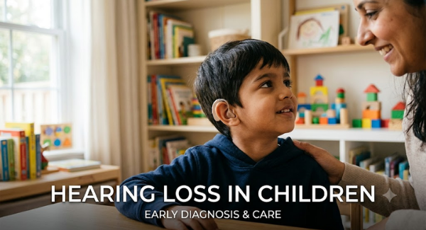
Children with permanent hearing loss who can benefit from amplification may be fitted with appropriately selected hearing aids. These devices amplify sound so that it becomes more accessible to the functioning auditory system. Regular audiological follow-up is important because children grow rapidly and their hearing needs can change.
Cochlear Implants
Children with severe-to-profound sensorineural hearing loss who receive insufficient benefit from hearing aids may be evaluated for cochlear implantation. Unlike a hearing aid, which amplifies sound, a cochlear implant bypasses damaged portions of the inner ear and electrically stimulates the auditory nerve. It does not restore normal hearing but can provide access to useful sound and speech information. Early implantation in appropriately selected children, followed by intensive rehabilitation, can support speech and language development.
Ayurveda Insight
In classical Ayurvedic literature, hearing loss in children is understood as Karnabadhirya, a condition categorised under the twenty-eight Karna Rogas and classified primarily as a Vata-predominant Nanatmaja Vyadhi. The auditory organ (Shrotrendriya) is considered an essential seat of Vata Dosha located in the head (Uttamanga), making it highly sensitive to physiological imbalances. The Ayurvedic pathophysiology (Samprapti) initiates when etiological factors—such as cold and dry diets, exposure to chilly winds, acoustic trauma, or neglected upper respiratory infections (Pratishyaya)—aggravate Prana Vayu and Kapha Dosha. These vitiated doshas travel upward into the head and pool within the sound-carrying channels (Shabdavaha Srotas). In pure Vataja presentations, the dry and degenerative nature of Vata causes atrophy of sensory nerve endings, closely mirroring sensorineural hearing loss. Conversely, when excess Kapha and unmetabolized metabolic waste (Ama) combine with Vata, they form an obstructive blockage (Avarana or Sanga) within the micro-channels, obstructing physical sound transmission in a manner analogous to conductive middle-ear effusion. Directing treatment toward pacifying Vata, clearing channel blockages through Srotoshodhana, and restoring micro-circulation via therapies like Karnapurana and Nasya helps protect residual hearing and support neural rehabilitation.
Role of Ayurvedic Ear Therapies
Traditional Ayurvedic procedures such as Karna Purana and the use of medicated oils are described for selected ear conditions. However, they should not be considered a universal treatment for childhood hearing loss. In particular, oil or other substances should not be placed into a child’s ear when there is ear discharge, suspected infection, tympanic membrane perforation, recent ear surgery, or an undiagnosed hearing problem. An ENT examination should precede any local ear procedure.
Herbal Remedies for Hearing Loss by Planet Ayurveda
Planet Ayurveda is a wellness brand dedicated to bringing traditional Ayurvedic principles into modern everyday life. Its range includes herbal and classical Ayurvedic formulations made with carefully selected natural ingredients. The brand focuses on supporting different aspects of wellness, including immunity, digestion, joint and bone health, skin care, and overall vitality. Planet Ayurveda combines traditional Ayurvedic knowledge with contemporary quality standards. Its formulations are designed to offer convenient and holistic support for healthy lifestyle practices. The brand aims to make Ayurveda more accessible and practical for people seeking natural wellness solutions. In this article, we will discuss some formulations for the management of Hearing loss in children. Let’s start!
- Ashwagandhaghan Vati
- Brahmi Chyawanprash
- Brahmi Vati (plain)
- Kumar Kalyan Ras
- Arvindasav
Herbal Remedies for Hearing Loss in Children


Product Description
1. Ashwagandhaghan Vati
Ashwagandhaghan Vati, prepared from concentrated Withania somnifera, is traditionally valued for its Rasayana (Rejuvenative), Balya (Strengthening), and Vata-pacifying actions. It may support healthy nerve function and help maintain the strength and nourishment of tissues involved in sensory processes. Its active withanolides have demonstrated antioxidant and neuroprotective properties, which may help protect cells from oxidative stress. Ashwagandha also supports healthy stress adaptation, energy levels, and overall neurological resilience. From an Ayurvedic perspective, its nourishing and Vata-balancing properties may be useful when weakness or Vata aggravation accompanies auditory difficulties in children.
Dosage: 1 tablet twice daily after meals with plain water.
2. Brahmi Chyawanprash
containing Brahmi along with Bilva, Agnimantha, Shyonaka, Kashmarya, Patala, Shalaparni and other herbs, may support children’s auditory and nervous-system health. Brahmi is traditionally considered Medhya Rasayana, supporting healthy nerve function and sensory processing. Its antioxidant properties may help protect neural tissues from oxidative stress. The nourishing Rasayana action of the formulation may support the tissues involved in sensory function during childhood growth. Its Vata-supportive properties are traditionally associated with maintaining normal nerve signalling and sensory coordination. Adequate nourishment and neurological resilience may be particularly relevant when a child has hearing difficulties. Thus, the formulation may provide holistic nutritional and neurological support for children with hearing impairment.
Dosage: ½ tsp twice daily after meals with Lukewarm milk/ water.
3. Brahmi Vati (Plain)
Brahmi Vati, containing Brahmi (Bacopa monnieri), Shankhapushpi (Convolvulus pluricaulis), Vacha (Acorus calamus), Maricha (Piper nigrum), Gojihva (Onosma bracteatum), Swarna Makshika Bhasma, and Ras Sindoor, is traditionally described as supporting Medhya, Vata-balancing, and nervous-system functions. Brahmi and Shankhapushpi are valued for supporting cognitive and neurological activity involved in sensory processing. Vacha is traditionally associated with maintaining clarity of sensory functions and Vata regulation. Maricha may support digestion and the bioavailability of accompanying herbal constituents. Swarna Makshika Bhasma is traditionally used for nourishment and strength of body tissues. These actions may provide supportive neurological and sensory nourishment relevant to children experiencing hearing difficulties.
Dosage: 1 tablet twice daily after meals with Plain water.
4. Kumar Kalyan Ras
Kumar Kalyan Ras, a classical Ayurvedic herbo-mineral formulation, commonly contains Swarna Bhasma, Mukta/Moti Bhasma, Abhrak Bhasma, Loha Bhasma, Swarna Makshika Bhasma, and Ras Sindoor, with variations between classical preparations and manufacturers. Traditionally, these ingredients are used for Balya (strengthening) and Rasayana (Rejuvenative) support, helping maintain strength, nourishment, and overall development in children. For hearing difficulties, its traditional role is better understood as supportive nourishment for the nervous and sensory systems rather than a direct action on the cochlea or auditory nerve. Some ingredients are traditionally considered Vata-supportive, which is relevant to the Ayurvedic understanding of sensory function.
Dosage: 1 tablet once daily after meals with water or as advised by a physician.
5. Arvindasav
Arvindasav, a classical Ayurvedic formulation containing Arjuna (Terminalia arjuna), Ashwagandha (Withania somnifera), Bala (Sida cordifolia), Shatavari (Asparagus racemosus), Mustak (Cyperus rotundus), and Draksha (Vitis vinifera), is traditionally used as a Balya (Strengthening) and Rasayan (Rejuvenative) preparation to support nourishment, strength, and healthy development in children. Its nourishing herbs may support overall neurological vitality and tissue health, which are relevant to normal sensory development. From an Ayurvedic perspective, maintaining balanced Vata and adequate Dhatu (Tissue) nourishment is considered important for sensory functions such as hearing. Arvindasav may therefore be considered supportive in a broader Ayurvedic approach when weakness or nutritional concerns accompany hearing difficulties. Childhood hearing loss, however, has diverse causes including genetic factors, infections, middle-ear disease, and birth-related complications. Early hearing assessment and timely intervention are important because untreated hearing loss can affect speech, language, learning, and social development.
Dosage: 2 tsp twice daily after meals with Lukewarm water.
Prevention of Childhood Hearing Loss
Prevention begins before birth and continues throughout childhood. Appropriate maternal healthcare, immunisation, prevention of congenital infections, safe use of medicines, and prompt management of ear diseases can reduce avoidable risks. Children should also be protected from unnecessary loud noise. Personal audio devices should be used at safe volumes, and prolonged exposure to loud environments should be minimised. Parents should never insert cotton buds, sharp objects, or home remedies into the ear canal. Recurrent ear infections and persistent hearing concerns should be evaluated rather than repeatedly managed at home. WHO’s 2026 World Hearing Day campaign specifically emphasises prevention, early identification, and access to hearing care through community and school settings.
Role of Parents and Teachers
Parents and teachers have a major role in recognising subtle hearing difficulties. A child who has difficulty hearing in a noisy classroom may appear inattentive or academically weak even though the underlying problem is auditory. Preferential seating, reducing background noise, speaking clearly, using visual material, and confirming that instructions have been understood can improve classroom participation. Families should also maintain regular audiological follow-up when hearing aids or cochlear implants are used. Emotional support is equally important. Children should be encouraged to communicate openly about hearing difficulties and should not be made to feel embarrassed about using hearing devices or alternative communication methods.
Conclusion
Childhood hearing loss has diverse causes and can influence communication, learning, social participation, and development. Conductive problems may sometimes be reversible, whereas sensorineural hearing loss may require long-term amplification, cochlear implantation, or other communication support. The most important principles are early screening, accurate diagnosis, timely intervention, rehabilitation, and continuous developmental support. WHO emphasises that prevention and early care can substantially reduce the impact of childhood hearing loss. From an Ayurvedic perspective, childhood ear disorders can be understood within the framework of Karna Roga, Vata Dosha, Shravana Indriya, Agni, and individualised health assessment. Ayurvedic dietary and lifestyle measures may provide supportive care, but they should be integrated responsibly with evidence-based audiological and ENT management. Ultimately, the objective is not merely to improve hearing measurements but to ensure that every child has the best possible opportunity to communicate, learn, participate, and develop with confidence.