Category: Brain Tumor

Common Signs of Oral Cancer You Should Never Ignore – Causes, Symptoms, Care & Treatment

Abstract

Oral cancer is the second most common cancer affecting the head and neck region. It can develop in any part of the oral cavity, including the tongue, lips, cheeks, floor of the mouth, and palate. It may also spread to the pharynx, throat, and tonsils. Oral cancer presents with a variety of signs and symptoms and can cause significant discomfort while talking, eating, or swallowing. The disease progresses through different stages, and many people remain undiagnosed until it reaches an advanced stage. A healthcare provider confirms the diagnosis through an oral examination and diagnostic tests such as a biopsy. Treatment options include surgery, radiotherapy, or chemotherapy, depending on the stage and type of cancer, as well as the patient’s overall health.
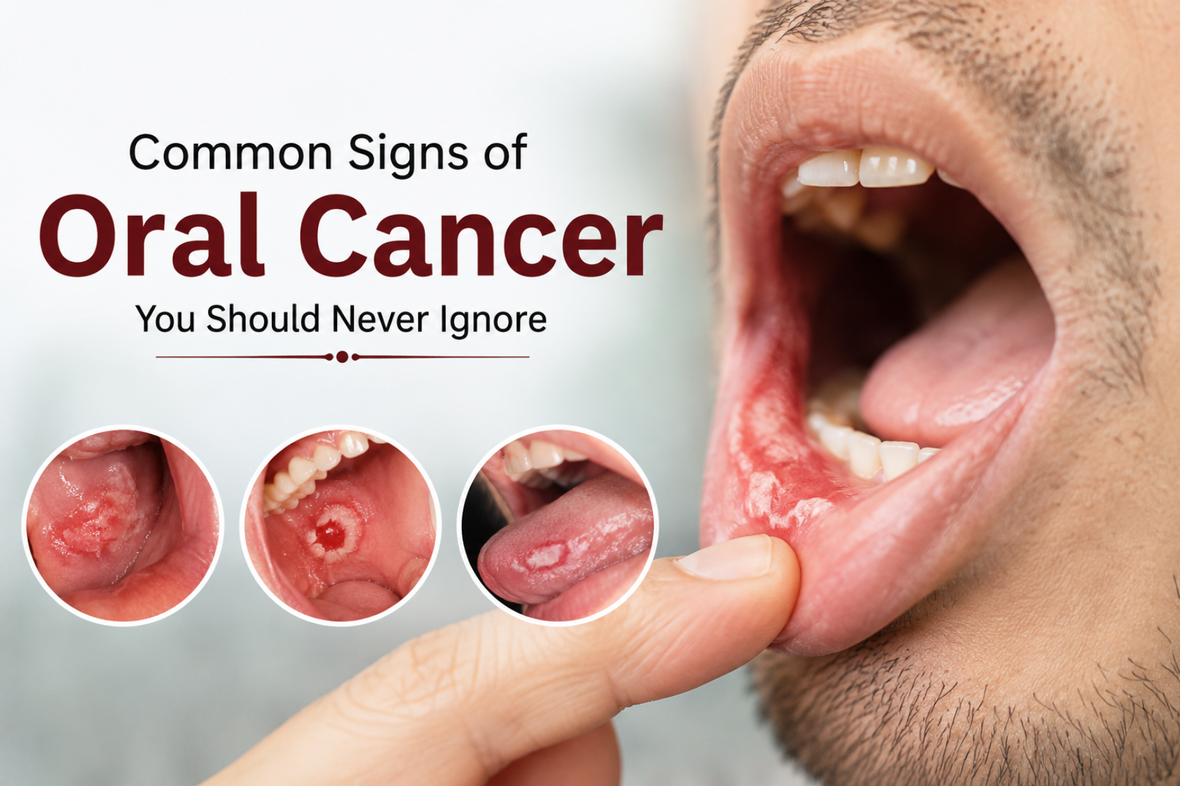
Common Signs of Oral Cancer

Statistics for Oral Cancer

  • In India, there are around 70000-77000 new cases of oral cancers each year.
  • The death rate with oral cancer is most common in India with 52000 cancer patients dying annually.
  • The average age of oral cancer prevalence is 64 years and 20% of the cases are under 55 years of age.
  • Oral cancer accounts for about 5% of all cancers worldwide.
  • Every hour, about five people die from oral cancer, reflecting its high mortality rate.

Introduction

Oral cancer is a broad term used to describe cancers that affect different parts of the oral cavity. Early signs may resemble common conditions such as white patches, mouth ulcers, or bleeding gums, making them easy to overlook. Oral cancer has the potential to spread to nearby structures in the head and neck region. It is more common in men than in women. The disease may also affect the oropharynx, including the palate, the posterior part of the tongue, and the middle part of the throat.

Different anatomical sites of Oral Cancer

  • Gums
  • Lips
  • Floor of mouth
  • Anterior part of tongue
  • Cheeks
  • Area behind the third molar tooth.

Types of Oral Cancer

There are several types of oral cancer based upon their location in the mouth. These are:

  • Squamous cell carcinoma – It affects some parts of the tongue, buccal mucosa, gingiva and lower lip.
  • Lymphoma – It affects the posterior area of the oral cavity, tonsillar region and base of the tongue.
  • Verrucous carcinoma – It affects the inner lining of cheeks, gingiva or the tongue.
  • Mucoepidermoid carcinoma – It is a tumor of the parotid salivary gland.
  • Adenoid cystic carcinoma – It is a salivary gland tumor
  • Melanoma – It affects the hard palate and upper gingiva.
  • Sarcoma – It affects the jaw bone and the buccal soft tissues.

Pathophysiology and risk factors of Oral Cancer

Grow and multiply uncontrollably. Most oral cancers arise from squamous cells, which are thin, flat cells that line the lips and the inside of the oral cavity. Changes or mutations in the DNA of these cells disrupt the normal regulation of cell growth and programmed cell death. As a result, abnormal cells continue to multiply while healthy cells die. Over time, these abnormal cells accumulate to form a mass known as a tumor. The tumor can invade nearby healthy tissues, and some cancer cells may eventually spread to distant parts of the body through a process known as metastasis.

Since there is no single cause of oral cancer, several risk factors contribute to its development. These include:

  • Alcohol: Excessive and frequent alcohol consumption increases the risk of oral cancer.
  • Smoking and tobacco use: Tobacco products such as cigarettes, cigars, pipes, chewing tobacco, betel nut, and snuff significantly increase the risk.
  • Ultraviolet (UV) radiation: Prolonged exposure to sunlight or tanning lamps increases the risk of lip cancer.
  • Human papillomavirus (HPV): HPV is transmitted through sexual contact. Although it often causes no symptoms, certain strains can alter cellular DNA and contribute to oral cancer.
  • Weak immunity and chronic stress: A weakened immune system due to medications, organ transplantation, HIV infection, or chronic stress may increase susceptibility to oral cancer.
  • Age: People aged 65–69 years are at a higher risk of developing oral cancer.
  • Family history: Individuals with a family history of cancer may have an increased risk because of shared genetic, environmental, and lifestyle factors.

Seven Common Signs of Oral Cancer that Should Not be Ignored

Recognizing the early signs of oral cancer can improve the chances of successful treatment. Early-stage oral cancer is often highly treatable. Therefore, it is important to seek medical evaluation if any of the following symptoms persist:

  • Persistent mouth sores: Mouth sores caused by accidental cheek biting, tongue biting, braces, or minor infections usually heal within one to two weeks. However, sores that persist for more than 14 days should be evaluated, as they may indicate a precancerous or cancerous condition. Initially, these sores may be painless but can later become painful or bleed.
  • Lumps in the mouth and neck: Any unexplained lump or thickened tissue in the mouth or neck that persists without an obvious cause should not be ignored. These lumps may be noticed while brushing, speaking, or chewing.
  • Red or white patches: White patches (leukoplakia) and red patches (erythroplakia) result from abnormal cell growth and are considered precancerous lesions. Red patches are particularly concerning because they have a higher likelihood of becoming cancerous. Mixed red-and-white patches may also occur.
  • Difficulty in jaw movements and swallowing: Progressive difficulty in opening the mouth or swallowing may indicate oral cancer. Pain while swallowing food or liquids and unintentional weight loss due to reduced food intake are common associated symptoms. Jaw stiffness may also contribute to temporomandibular joint (TMJ) disorders.
  • Persistent pain in ear:  Long-lasting ear pain without an obvious ear infection may be referred to as pain from a tumor in the oral cavity or throat and should be medically evaluated.
  • Changes in voice or hoarseness: Persistent hoarseness, a strained or raspy voice, or difficulty speaking should not be ignored, particularly if accompanied by mouth sores, difficulty swallowing, or unexplained weight loss.
  • Bleeding in the mouth: Unexplained bleeding from the mouth, especially while brushing the teeth or blood in the saliva without an obvious cause, requires prompt medical evaluation.

Diagnosis and staging of Oral Cancer

Anyone experiencing symptoms suggestive of oral cancer should consult a healthcare provider for evaluation. Early diagnosis can prevent disease progression and improve treatment outcomes. Several diagnostic tests are used to confirm the diagnosis:

  • Incisional biopsy: The doctor removes a small sample of tissue for examination. If the suspicious area appears concerning, they may also remove the entire lesion along with a small margin of surrounding healthy tissue.
  • Oral brush biopsy: Cells are collected by gently brushing the affected area with a specialized brush.
  • Fine-needle aspiration (FNA): A thin, hollow needle attached to a special syringe is used to collect a sample of cells from a lump in the neck or an enlarged lymph node for laboratory examination.
  • Endoscopy: The doctor examines the throat using a flexible tube fitted with a tiny camera. If any abnormal or suspicious areas are identified, tissue samples may be collected for further analysis.
  • HPV testing: In cancers involving the throat or the back of the tongue, tumor cells may be tested for HPV, as the presence of the virus influences cancer staging.
  • Imaging tests (CT, MRI, and PET/CT): CT and MRI scans help determine the size and location of the tumor and assess whether the cancer has spread to nearby lymph nodes or other parts of the body.

Staging of Oral Cancer

Oral cancers are classified on the basis of size of tumor, lymph node involvement and its spread. TNM staging is done for this purpose. T denotes tumor size, N denotes nodal involvement and M denotes metastasis.

T = Primary tumor

  • Tx – Tumor cannot be detected
  • Tis – Carcinoma in-situ
  • T1 – Size of tumor is less than or equal to 2cm
  • T2 – Size of tumor is more than 2cm
  • T3 – Size of tumor is greater than 2cm or equal to 4cm
  • T4 -Size of tumor is greater than 4cm

N = Regional lymph node

  • NX Nodes cannot be detected
  • N0 No spread to lymph nodes.
  • N1 Spread to one lymph node on the same side, with size 3 cm or less
  • N2 Spread to one large or several  lymph nodes on the same or opposite side
  • N3 Spread to a node larger than 6 cm, or there is multi-node involvement

M = Distant metastasis

  • M0 – No spread
  • M1 – Spread to other parts of body

There are different stages of oral cancer which are as follows:

  • Stage 0 – Carcinoma in-situ : Tis, N0, M0
  • Stage 1 – T1, N0, M0
  • Stage 2 – T2, N0, M0
  • Stage 3 – T3, N0, M0 or T1/T2/T3, N1, M0
  • Stage 4a – T4a, (N0, N1), M0 or (T1, T2, T3, T4a), N2, M0
  • Stage 4b – (Any T), N3, M0 or T4b, (Any N), M0
  • Stage 4c – (Any T), (Any N), M1

Conclusion

Oral cancer is a serious but largely preventable disease that requires early recognition and timely treatment. The main causes include tobacco, alcohol, HPV infection, and long-term sun exposure. Diagnosis is confirmed through clinical examination, biopsy, and imaging, while staging helps determine the extent of the disease and guides treatment planning. Early diagnosis greatly improves prognosis and survival. Raising awareness, reducing risk factors, and encouraging regular oral examinations are essential for improving survival and quality of life.
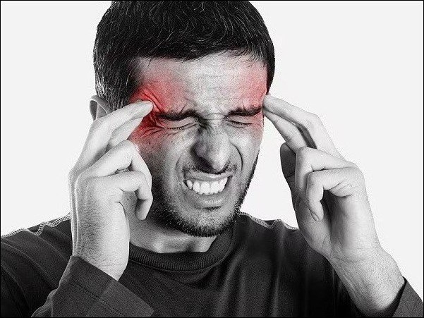
Leptomeningeal Enhancement Symptoms And Treatment By Ayurveda

Abstract

Meninges are the membranes that envelop and protect the brain and spinal cord. These meninges are the dura mater, arachnoid mater, and pia mater. The main function of the meninges is to protect the CNS (central nervous system). Sometimes there are conditions that these meninges become inflamed or increase in size due to major infections and other serious issues. The increase in the size of these meninges is known as a leptomeningeal enhancement. In the text below, we shall discuss this condition in detail and see its management in the contemporary system of medicine as well as in the traditional system of Ayurvedic medicine. Let’s see !!

Leptomeningeal Enhancement

Introduction

The increase in the brain surface is called pial or pial-arachnoid enhancement and is generally known as a leptomeningeal enhancement. Thin arachnoid film is connected or attached to the inner surface of the dura mater, the pachymeningeal pattern of enhancement can likewise be depicted as a dural-arachnoid enhancement. This enhancement can be due to a number of conditions from infection to tumour. Therefore, the management is needed as soon as leptomeningeal enhancement is detected. Sometimes the patient may be asymptomatic and and sometimes associated with non specific neurological deficits. The conditions can be related to the tridosha imbalance in Ayurvedic context. Now let’s see the causes, classification, features, management and ayurvedic aspect of the condition. 

Causes of Leptomeningeal enhancement

  • Amoebic meningoencephalitis
  • Encephalitis
  • Granulomatous conditions
  • Haemorrhage
  • Tumours/cancers
  • Post operative infection
  • Leptomeningeal carcinomatosis
  • Vasculitis
  • Neurosarcoidosis
  • Neurosyphilis
  • Sturge-Weber syndrome etc

Signs and symptoms

  • Nausea and vomiting
  • Headaches
  • Dizziness
  • Double vision
  • Difficult speaking
  • Swallowing difficulty
  • Pain in arms and legs
  • Loss of bladder and bowel control
  • Seizures
  • Weakness
  • Slack of coordination in the arms and legs
  • Fever
  • Hearing loss
  • Back pain

Investigations 

  • Lumbar puncture
  • CT Scan
  • Magnetic Resonance Imaging (MRI)

Management of Leptomeningeal enhancement 

  • Radiation
  • Intrathecal-thecal chemotherapy
  • Immunotherapy
  • Antineoplastic agents
  • Tyrosine kinase inhibitors
  • Antimetabolites
  • Surgical intervention

Ayurvedic aspect and management of Leptomeningeal Enhancement 

Ayurveda, the old age science continuously helps mankind with its marvellous results. Ayurveda revolves around the three basic principles or dosha which are vata, pitta and kapha. There are seven dhatus as well. Any kind of vitiation in these doshas and  dhatus results in disease and within normal range leads to a healthy body, mind and soul. The condition of Leptomeningeal enhancement which we are discussing can not be correlated directly to any disease mentioned in the ayurvedic texts  but according to the symptoms, and findings this can ideally find its place in the disorders referred to as tridoshaja roga.  

Let’s understand how the three dosha are involved in this disease. As the condition can be due to many causative factors which results in enhancement of the meninges. Therefore, the enhancement is the features of vitiated kapha dosha. Further if the condition is due to inflammation caused by infectious agents then this comes out to be a condition due to pitta dosha dominance. And the features such as diplopia, seizures and other neurological features are related to the vitiated vata dosha. Every case of this disease has all the tridosha vitiation and hence regarded as tridoshaja vyadhi. And features of the conditions depend on the dosha predominance.

There are many herbs that are effective correcting the vitiated dosha such as bilva (Aegle marmelos), patla (Stereospermum suaveolens), gambhari (Gmelina arborea), amla (Embelica officinalis), bibhitaki (Terminalia billerica), haritaki (Terminalia chebula), pippali (Piper longum), kachnaar (Bauhinia variegata), prishniparni (Uraria picta), yashtimadhu (Glycyrrhiza glabra), ashwagandha (Withania somnifera), brahmi (Bacopa monnieri), guduchi (Tinospora cordifolia), haridra (Curcuma longa),ginger (Zingiber officinale), marich (Piper nigrum), musli (Chlorophytum borivilianum) and various others.

Kanchnaar guggul, Ras Sindur, Haridra khand, Ashwagandha ghan vati, Chandraprabha vati, Shallaki and various other formulations are available that are effective in managing the condition of Leptomeningeal enhancement.

Herbal Remedies for Leptomeningeal enhancement by Planet Ayurveda

Planet Ayurveda is an eminent GMP certified, ISO 9001:2015 certified and US-FDA registered Ayurvedic Company, which adheres to the objective of manufacturing quality Ayurvedic products as mentioned in a number of ancient texts of Ayurveda. Formulations are manufactured without additives and preservatives and other kinds of chemicals or artificial flavouring agents which can have a number of side effects on the human body. Products manufactured at Planet Ayurveda are pure and devoid of any kind of adulteration. The products are purely organic, vegetarian, gluten free and halal certified. Planet Ayurveda presents its product that effectively manages the condition of Leptomeningeal enhancement. 

Following is the list of formulations that are helpful in managing Leptomeningeal Enhancement 

Product List 

  1.  Neurogenie Capsules 
  2.  Yogendra Ras
  3.  Pitta Balance
  4.  Kanchnaar Guggul
  5.  Boswellia Curcumin
  6.  Neuro Plan Syrup
  7.  Musli Strenght Capsules
  8.  Vikramprash
    Herbal Remedies For Leptomeningeal Enhancement 

Herbal Remedies For Leptomeningeal Enhancement 

Product Description

1. Neurogenie Capsules 

Neurogenie capsules are composed of  the herbs which are medhya and promote and improve neurological functions as mentioned in the Ayurvedic texts. The formulation is filled with the properties of herbs brahmi (Bacopa monnieri) and ashwagandha (Withania somnifera). As these are beneficial in promoting muscle health and nerve conduction and their coordination. The herbs in the formulation are vata pacifiers therefore manage the disorders of vata vitiation and restrict and reverse the abnormal growth of the meninges and support the  healthy nervous tissue. With these properties, Neurogenie capsules help in Leptomeningeal enhancement.

Dosage : One capsule twice a day with plain water after meals.

2. Yogendra Ras

Yogendra Ras is a classical Ayurvedic formulation. It is a mineral formulation which is helpful in various health conditions.  It is composed of loha bhasma (iron calx), shuddha parada (purified mercury), gandhak (purified sulphur), ras sindoor, vanga bhasma (tin calx), shuddha mukta (purified pearls), swarna bhasma (gold calx) and abhrak bhasma (purified silica). Yogendra Ras increases the efficacy of other medicines when taken along with it. Yogendra Ras provides strength to the nerves and muscle and helps in good nervous tissue and health. Therefore, Yogendra Ras is beneficial in Leptomeningeal enhancement.

Dosage : One tablet twice a day.

3. Pitta Balance

Pitta balance is a product of Planet Ayurveda that balances the pitta dosha. It is a herbomineral formulation which contains jaharMohra, kaharawa, akik, mukta, giloy satva (Tinospora cordifolia). As it manages the pitta vitiation, thus helps in symptoms associated with vitiated pitta. It reduces the inflammation thus helps in Leptomeningeal enhancement.

 Dosage : One capsule twice a day with plain water after meals.

4. Kanchnaar Guggul

Kanchnaar guggul is beneficial in Leptomeningeal enhancement as it reduces the size of the abnormal enhancement of the meninges. It contains the herbs that are known to reduce the abnormal sizes of the tissues and reduce pain. Ingredients of the formulation include kachnar bark (Bauhinia variegata), amalaki (Emblica officinale), haritaki (Terminalia chebula), bibhitaki (Terminalia billerica), ginger (Zingiber officinale), marich (Piper nigrum), pippali (Piper longum), varuna bark (Crataeva religiosa), tejapatra (Cinnamomum tamala), guggul (Commiphora mukul) etc. The formulation reduces the size of the neoplastic type of Leptomeningeal enhancement. Thus it is a magical formulation that Ayurveda has in uncontrolled and abnormal tissue growths.

Dosage : Two tablets twice a day with lukewarm water.

5. Boswellia Curcumin

Boswellia Curcumin is a product of Planet Ayurveda which is enriched with the goodness of shallaki (Boswellia aristata) and haridra (Curcuma longa). The herbs have healing properties and also possess analgesic effects which makes it an effective formulation in the Leptomeningeal enhancement. It regresses the inflammation, heals the damaged tissue and also alleviates the pain and reduces the abnormal size or enhancement of the meninges.

Dosage : One capsule twice a day.

6. Neuro Plan Syrup

Neuro Plan Syrup is a remarkable formulation for increasing neurological health and managing all the neurological conditions. The syrup contains a variety of herbs which assuredly manages Leptomeningeal enhancement . The syrup is enriched with the properties of mandukaparni (Centella asiatica), jyotishmati (Celastrus paniculata), madhuyashti (Glycyrrhiza glabra), pippali (Piper longum), maricha (Piper nigrum), ashwagandha (Withania somnifera), shankhpushpi (Convolvulus pluricaulis), brahmi (Bacopa monnieri) etc. All the herbs are known to have medhya properties which means these are helpful in maintaining a good neurological health. The herbs heal the damage of the nerves. It improves motor function. It relieves symptoms of vitiated vata dosha. Therefore, by pacifying the vata dosha, it helps in alleviating the seizure-like symptoms of Leptomeningeal enhancement.

Dosage : Two tsp twice a day.

7. Musli Strength Capsules

Musli Strength Capsules are enriched with properties of gokshura (Tribulus terrestris) and safed musli (Chlorophytum borivilianum). Both the herbs pacify vata dosha. As the name suggests Musli Strength Capsules strengthens the muscles and improves neurological health. The capsules help in the neoplastic condition of the nervous tissues and heals the damaged neurons. Therefore, Musli Strength Capsules are helpful in managing Leptomeningeal enhancement.

Dosage : One capsule twice a day.

8. Vikramprash

Vikramprash is a preparation of Planet Ayurveda which is available in jam form. Vikramprash is filled with the herbal properties of  Bilva (Aegle marmelos), Agnimantha (Premna mucronata), Syonak (Oroxylum indicum), Patla (Stereospermum suaveolens), Pippali (Piper longum), Gokshura (Tribulus terrestris), Jivanti (Leptadenia reticulata), Utpal (Nymphaea alba), Musta (Cyperus rotundus) and many other herbs. Drugs that are present in the formulation promotes the wellbeing of  a person because it maintains the dosha levels within normal limits. Antioxidants present in the medicine slow down the ageing process because it is rich in nutrients that rejuvenates body cells. Being fulfilled with the properties of rasayana, Vikramprash helps in boosting immunity that further benefits in disorders of autoimmune, age related and neoplastic origin. With amazing and tridosha balancing  properties, Vikramprash helps in management of Leptomeningeal enhancement.

Dosage : One spoon daily

CONTACT  PLANET AYURVEDA  TO PROVIDE YOU WITH THE    COSTING / ORDERING AND DELIVERY INFORMATION AT – COSTING.PLANETAYURVEDA@GMAIL.COM OR CALL AT +91-172-5214040

Conclusion 

As we discussed Leptomeningeal enhancement, we have come to know that leptomeningeal enhancement is an abnormal growth or enhancement of the meninges that can occur due to a number of factors including infections, tumours, encephalitis, granulomatous conditions, haemorrhage etc. Leptomeningeal enhancement may stay asymptomatic in some cases but in some cases it may lead to a number of troublesome features. In the modern system of medicine, there is no proper management available but chemotherapy and radiation therapy are some of the main management therapies that are performed which does not provide positive effects efficiently but causes a number of side effects which can hamper other functions of the body. Whereas Ayurvedic management is based on correcting the dosha imbalance that causes the disorder and also manages the symptoms impeccably. Further herbs are available that beautifully contain the symptoms and disease to the maximum. Hence, preferring Ayurveda over the side effect causing therapies will eventually provide promising results.