ELECTROCARDIOGRAM (ECG / EKG): WHAT IS IT? WHY IS IT DONE? HOW CAN AYURVEDA HELP SUPPORT HEART RHYTHM?
ABSTRACT
An Electrocardiogram (ECG or EKG) is a non-invasive diagnostic test that records the electrical activity of the heart to assess its rhythm, rate, and overall function. It is commonly used to detect arrhythmias, myocardial infarction (heart attack), conduction abnormalities, electrolyte imbalances, and structural cardiac changes. The test is quick, painless, and widely performed in outpatient clinics, emergency departments, and hospitals. By analyzing wave patterns such as the P wave, QRS complex, and T wave, healthcare professionals can identify irregularities in cardiac electrical conduction. Early detection through ECG plays a crucial role in preventing serious complications. While modern medicine focuses on diagnosis and acute management, Ayurveda offers supportive measures aimed at balancing doshas, strengthening cardiac function, reducing stress, and promoting overall heart health.
WHAT IS AN ELECTROCARDIOGRAM (ECG)?
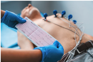
An Electrocardiogram is a test that measures the electrical impulses generated by the heart as it contracts and relaxes. These electrical signals are recorded through electrodes placed on the chest, arms, and legs, producing a graphical tracing on paper or a digital monitor.
The ECG tracing shows different waves:
- P wave: Atrial depolarization
- QRS complex: Ventricular depolarization
- T wave: Ventricular repolarization
Any abnormality in these waves may indicate a heart problem.
WHY IS ECG DONE?
An ECG is performed to:
- Detect irregular heart rhythms (arrhythmias)
- Diagnose heart attack (myocardial infarction)
- Evaluate chest pain
- Assess palpitations
- Monitor heart disease
- Check pacemaker function
- Detect electrolyte imbalances
- Evaluate effects of certain medications
It is often the first-line investigation in suspected cardiac conditions.
TYPES OF ECG
- Resting ECG: Done while lying down.
- Stress ECG (Treadmill Test): Performed during exercise.
- Holter Monitoring: Continuous ECG recording for 24–48 hours.
- Event Recorder: Used for intermittent symptoms over longer periods.
WHAT CONDITIONS CAN ECG DETECT?
- Atrial fibrillation
- Ventricular tachycardia
- Heart block
- Myocardial infarction
- Left ventricular hypertrophy
- Electrolyte disturbances (e.g., potassium imbalance)
- Pericarditis
HOW IS THE PROCEDURE DONE?
Before the test begins, the patient is asked to lie down comfortably on an examination table in a relaxed position. The chest area may be cleaned, and in some cases, small areas of hair are shaved to ensure proper electrode contact. About 10 small adhesive electrodes are placed on specific locations on the chest, arms, and legs. These electrodes are connected to the ECG machine through lead wires. The patient is instructed to remain still, breathe normally, and avoid talking during the recording, as movement can interfere with the tracing. The machine then records the heart’s electrical activity for a few seconds, producing a waveform printout or digital report. The entire procedure typically takes 5–10 minutes. It is completely painless and safe, as it only records electrical impulses and does not send electricity into the body.
LIMITATIONS OF ECG
Although an ECG is a valuable and widely used diagnostic tool, it has certain limitations. A normal ECG does not completely rule out heart disease. Some conditions, such as early coronary artery disease or mild structural abnormalities, may not produce visible changes on a resting ECG. Therefore, a patient may still have underlying cardiac issues despite a normal report. Additionally, ECG records the heart’s electrical activity only during the few seconds of testing. Intermittent rhythm disorders, such as occasional atrial fibrillation or episodic tachycardia, may not occur during the recording and can be missed. In such cases, longer monitoring methods like Holter monitoring or event recorders are more useful. ECG also provides limited information about the heart’s structure and pumping efficiency. It cannot directly visualize heart valves, chambers, or blood flow. For a more detailed evaluation, additional tests such as echocardiography, stress testing, cardiac CT, or coronary angiography may be required. Therefore, ECG is often the first step in cardiac assessment, but not always the final diagnostic tool.
AYURVEDIC VIEW ON COMMON ECG-DETECTED CONDITIONS
Atrial Fibrillation
In Ayurveda, atrial fibrillation may be correlated with Vataja Hridroga, where aggravated Vata disturbs the normal rhythmic movement of the heart. Irregular, rapid, and chaotic heartbeat reflects Vata vitiation affecting Prana Vayu and Vyana Vayu. Stress, anxiety, aging, excessive dryness, and irregular lifestyle are considered contributing factors. Management focuses on Vata pacification, calming the mind, and strengthening Hridaya (heart tissue).
Ventricular Tachycardia
Ventricular tachycardia may be interpreted as aggravated Vata associated with Pitta, leading to excessive speed and force in cardiac activity. Increased heat, irritability, and overstimulation may reflect Pitta involvement. It can be considered a severe disturbance of cardiac rhythm. Ayurveda emphasizes stabilization of Vata, cooling Pitta if aggravated, and supporting Ojas (Vital Nectar of life) to maintain cardiac stability.
Heart Blockage
Heart blockage may be viewed as obstruction (Avarana) in the channels (Srotas) responsible for conduction of impulses. When Vata’s movement is obstructed by Kapha or structural imbalance, signal transmission slows down. This condition correlates with impaired Prana and Vyana Vayu coordination. Ayurvedic perspective focuses on removing obstruction, improving Srotas patency, and restoring balanced Vata flow.
Myocardial Infarction
Myocardial infarction can be correlated with chronic Hridroga involving Kapha-Pitta imbalance and obstruction of Rasavaha and Raktavaha Srotas (Channels of Circulatory fluids and blood tissue). Accumulation of Ama (metabolic toxins) and Meda (lipid imbalance) may block circulation, leading to tissue damage. Aggravated Pitta may contribute to inflammation and burning sensation. Management in Ayurveda emphasizes Ama pachana, improving circulation, and strengthening heart tissue.
Left Ventricular Hypertrophy
Left ventricular hypertrophy may be interpreted as long-standing pressure imbalance leading to structural adaptation of the heart. From an Ayurvedic view, chronic Vata aggravation with Kapha association may cause thickening or enlargement. Persistent hypertension (often linked with Vata-Pitta imbalance) may contribute. Treatment principles include regulating blood pressure naturally, balancing doshas, and maintaining proper circulation.
Electrolyte Disturbances (e.g., Potassium Imbalance)
Electrolyte imbalance may correlate with disturbance in body fluid balance and Agni (Digestive fire) dysfunction. Improper diet, dehydration, or metabolic disturbances can impair Rasa Dhatu quality. Vata becomes unstable when mineral balance is disturbed, potentially affecting heart rhythm. Ayurveda emphasizes proper hydration, balanced diet, Agni correction, and maintaining Dhatu (Body Tissues) equilibrium.
Pericarditis
Pericarditis may be correlated with inflammatory conditions of the heart covering, often involving aggravated Pitta and Rakta Dushti (vitiated blood tissue). Symptoms like chest pain and burning sensation indicate Pitta dominance, while Vata may contribute to sharp pain. Ayurvedic management focuses on Pitta pacification, reducing inflammation, detoxifying Rakta (Blood Tissue), and supporting cardiac strength.
AYURVEDIC HERBS TO SUPPORT COMMON ECG-DETECTED CONDITIONS
1. Arjuna (Terminalia arjuna)
Arjuna is described as a potent Hridya (cardiotonic) that nourishes and strengthens the heart muscle (Mamsa Dhatu of Hridaya). In atrial fibrillation and ventricular tachycardia, where Vata disturbs the normal rhythm, Arjuna helps stabilize cardiac activity by supporting proper contraction and relaxation. In myocardial infarction and LVH, it assists in improving coronary circulation and oxygen delivery. Its Kashaya Rasa helps reduce excess Pitta and Rakta Dushti involved in inflammatory states like pericarditis.
2. Ashwagandha (Withania somnifera)
Ashwagandha primarily pacifies Vata and strengthens Ojas. Since many rhythm disorders are triggered by stress and autonomic imbalance, this herb supports parasympathetic stability and reduces sympathetic overdrive. In chronic hypertension contributing to LVH, its adaptogenic effect helps regulate stress hormones. It also improves overall cardiac resilience during recovery phases after ischemic injury.
3. Pushkarmoola (Inula racemosa)
Pushkarmoola supports Prana Vayu and Vyana Vayu coordination. In heart block or ischemic conditions, where conduction or circulation is compromised, it helps maintain Srotas openness and oxygenation. It is traditionally indicated in Hridroga with breathlessness, suggesting its supportive role in improving cardiac output and relieving Kapha-related obstruction.
4. Jatamansi (Nardostachys jatamansi)
Jatamansi calms aggravated Vata and Pitta affecting the heart and mind connection. In arrhythmias triggered by emotional stress, anxiety, or inflammation, it promotes mental tranquility and reduces internal heat. Its antioxidant nature may support cellular protection in inflammatory conditions like pericarditis and electrolyte-related irritability.
5. Sarpagandha (Rauvolfia serpentina)
Sarpagandha influences autonomic nervous system regulation. In ventricular tachycardia or hypertension-induced LVH, it helps reduce excessive cardiac excitability and blood pressure. By calming Vata-Pitta overactivity, it supports controlled heart rate. However, due to potent alkaloids, careful medical supervision is essential.
6. Guggulu (Commiphora mukul)
Guggulu acts on Meda Dhatu and clears Ama accumulation. In myocardial infarction or atherosclerotic tendencies, it supports lipid regulation and improves circulation. By maintaining Srotas patency, it helps prevent Kapha-related obstruction contributing to ischemic heart disease.
7. Brahmi (Bacopa monnieri)
Brahmi enhances parasympathetic tone and reduces stress-induced palpitations. It supports mental clarity and stabilizes Prana Vayu, thereby indirectly assisting in rhythm regulation, especially in atrial fibrillation associated with anxiety.
8. Shankhpushpi (Convolvulus pluricaulis)
Shankhpushpi works as a nervine tonic that calms Vata-driven irregular heartbeat. It supports emotional balance, reduces restlessness, and helps regulate neuro-cardiac signaling pathways involved in rhythm disturbances.
CONCLUSION
An Electrocardiogram (ECG / EKG) is a fundamental diagnostic tool that plays a vital role in identifying rhythm disturbances, ischemic changes, conduction abnormalities, and other cardiac conditions at an early stage. It provides quick, safe, and valuable insight into the electrical functioning of the heart, allowing timely medical intervention and prevention of serious complications. While modern cardiology focuses on accurate diagnosis and emergency management, Ayurveda offers a supportive and holistic perspective by addressing underlying doshic imbalance, improving circulation, strengthening cardiac tissue, and enhancing Ojas. Through appropriate lifestyle modifications, dietary regulation, stress management, and carefully selected herbs under professional supervision, Ayurveda can complement conventional care and contribute to long-term heart rhythm stability and overall cardiovascular well-being.
