What Are Ketosis And Ketoacidosis? Its Ayurvedic Aspect
Abstract
Ketosis and ketoacidosis represent two distinct metabolic states characterised by the accumulation of ketone bodies in the bloodstream. While nutritional ketosis is a regulated, physiological adaptation to low carbohydrate availability where ketone bodies serve as an efficient alternative fuel source, ketoacidosis—most commonly Diabetic Ketoacidosis (DKA)—is an uncontrolled, life-threatening metabolic crisis marked by severe hyperglycemia, hyperketonemia, and metabolic acidosis. Understanding the biochemical divergence between these two states is essential for clinical management and metabolic health optimisation. This article details the definitions, metabolic pathways, triggers, clinical manifestations, diagnostic markers, and modern management of both ketosis and ketoacidosis. Furthermore, it integrates an Ayurvedic analysis, examining nutritional ketosis through the lens of Langhana (depletion/fasting therapies) and Medodhatu (fat tissue) metabolism, while mapping ketoacidosis to severe Agni Mandya (derangement of digestive/metabolic fire), Vata-Pitta exacerbation, and Prameha Upadrava (advanced complications of metabolic/diabetic disorders).
Introduction
The human body continuously changes its energy source according to nutritional availability and hormonal signals. After eating, glucose is an important energy substrate. During fasting or significant carbohydrate restriction, insulin levels decrease, and the body increasingly mobilises stored fat. The liver converts fatty acids into ketone bodies, which can serve as an energy source for several tissues, including the brain. This process is known as ketosis. In an otherwise healthy person, nutritional ketosis is generally controlled by hormonal mechanisms and does not usually produce severe metabolic acidosis. Ketoacidosis is fundamentally different. It represents a pathological state in which ketone production becomes excessive and contributes to an accumulation of acids in the blood. Diabetic ketoacidosis is most commonly associated with inadequate insulin activity and increased counter-regulatory hormones. The resulting metabolic abnormalities include ketonemia, metabolic acidosis, dehydration, and electrolyte disturbances. Understanding the difference is especially important because the word “ketosis” is sometimes used loosely to describe both normal dietary ketosis and dangerous ketoacidosis.
What Is Ketosis?
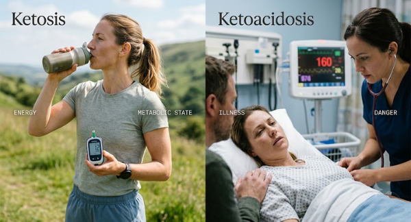
Ketosis is a metabolic state in which the body relies more heavily on fat as an energy source and produces increased quantities of ketone bodies. The principal ketone bodies are beta-hydroxybutyrate, acetoacetate, and acetone. Ketosis develops when carbohydrate availability or insulin-mediated glucose utilisation decreases sufficiently to increase fat mobilisation. Common physiological circumstances include overnight fasting, prolonged fasting, extended exercise, and carbohydrate-restricted diets. During nutritional ketosis, insulin remains present at levels sufficient to restrain excessive lipolysis and ketone production. Nutritional Ketosis is a physiological state wherein circulating blood ketone levels (specifically beta-hydroxybutyrate) range between 0.5 mmol/L and 3.0 mmol/L. Serum pH remains strictly within the normal physiological range (7.35 to 7.45). It occurs when dietary carbohydrate intake drops below the threshold required for oxaloacetate regeneration in the Krebs cycle, forcing the liver to convert free fatty acids into ketones.
What Is Ketoacidosis?
Ketoacidosis occurs when ketone production exceeds the body’s ability to utilise and eliminate ketone bodies, resulting in accumulation of acidic compounds and a fall in blood pH. The most important clinical form is diabetic ketoacidosis (DKA). According to the 2024 international consensus report, DKA involves three major abnormalities: diabetes or significant hyperglycemia, elevated ketone concentrations, and metabolic acidosis. Ketoacidosis is a pathological state characterised by serum ketone levels typically exceeding 3.0 mmol/L (often rising above 10 to 20 mmol/L), accompanied by marked blood glucose elevation (typically >250 mg/dL, though euglycemic variants exist) and a reduction in arterial blood pH below 7.30 (and serum bicarbonate <18 mEq/L). It occurs primarily in Type 1 Diabetes Mellitus, advanced Type 2 Diabetes under physiological stress, or severe alcohol abuse. Ketoacidosis can also occur in other situations, including prolonged starvation, excessive alcohol consumption, pregnancy-associated metabolic stress, and certain medication-related states. These conditions have different underlying mechanisms and should not automatically be classified as diabetic ketoacidosis.
Ketosis Versus Ketoacidosis
The key difference is physiological regulation and acid-base status. In ordinary ketosis, increased fat utilisation leads to controlled ketone production without clinically significant metabolic acidosis. It represents an adaptation to altered fuel availability. In ketoacidosis, ketone production becomes excessive and is associated with metabolic acidosis. In DKA, insufficient insulin activity prevents effective glucose utilisation and removes the normal restraint on fat breakdown. At the same time, hormones such as glucagon, adrenaline, cortisol, and growth hormone increase, promoting glucose production and ketogenesis. Therefore, the presence of ketones alone does not establish ketoacidosis. Blood ketone concentration, acid-base measurements, glucose status, clinical symptoms, and the underlying cause must be considered together.
Biochemical Mechanisms and Causes
Mechanisms of Nutritional Ketosis
Under low carbohydrate conditions, insulin secretion decreases while glucagon levels rise in modest amounts. This hormonal shift activates hormone-sensitive lipase in adipose tissue, releasing free fatty acids into circulation. These fatty acids travel to the liver, entering the mitochondria for beta-oxidation. The resulting acetyl-CoA exceeds the processing capacity of the citric acid cycle due to oxaloacetate depletion. Excess acetyl-CoA is funnelled into ketogenesis. Insulin levels, though low, remain sufficient to prevent runaway lipolysis and preserve systemic acid-base homeostasis.
Causes of Nutritional Ketosis
Low-carbohydrate high-fat (ketogenic) diets, intermittent or prolonged therapeutic fasting, extended endurance exercise, and starvation.
Mechanisms of Ketoacidosis
In Diabetic Ketoacidosis (DKA), absolute or near-total absence of insulin prevents glucose uptake into cells. Sensing cellular starvation, the body releases massive surges of counter-regulatory hormones (glucagon, epinephrine, cortisol, and growth hormone). This triggers unrestrained lipolysis in adipose tissue, flooding the liver with fatty acids. Uninhibited hepatic ketogenesis produces immense quantities of acetoacetic acid and beta-hydroxybutyric acid, which dissociate into hydrogen ions. The accumulation of hydrogen ions exhausts the bicarbonate buffering capacity of the plasma, dropping systemic blood pH and precipitating severe metabolic acidosis.
Causes of Ketoacidosis
Undiagnosed Type 1 Diabetes, missed insulin doses, severe infection or sepsis, acute cardiovascular events, trauma, heavy alcohol consumption (Alcoholic Ketoacidosis), or the use of SGLT2 inhibitors (which can trigger euglycemic DKA).
Clinical Signs and Diagnostic Evaluation
Nutritional Ketosis
- Signs and Symptoms: Mild initial fatigue (“keto flu”), temporary alteration in bowel habits, transient headaches, mild dry mouth, increased thirst, fruity breath odour (due to volatile acetone elimination), heightened mental clarity, and decreased appetite.
- Diagnostic Markers: Blood beta-hydroxybutyrate between 0.5–3.0 mmol/L; normal blood glucose (70–100 mg/dL); normal arterial/venous pH (7.35–7.45); normal serum bicarbonate (22–28 mEq/L).
Ketoacidosis
- Signs and Symptoms: Rapid, deep breathing designed to blow off carbon dioxide and reduce acidity (Kussmaul respiration); strong fruity/sweet breath odour; severe nausea, vomiting, and diffuse abdominal pain; profound dehydration, dry mucous membranes, and decreased skin turgor; confusion, lethargy, cerebral oedema, and potential coma.
- Diagnostic Markers: Blood beta-hydroxybutyrate >3.0 mmol/L; blood glucose usually >250 mg/dL; arterial blood pH <7.30; serum bicarbonate <18 mEq/L; elevated anion gap (>12 mEq/L); presence of urinary ketones and glucosuria.
Modern Management of Ketoacidosis
DKA is treated as an acute medical emergency. The major therapeutic objectives are restoration of circulating volume, suppression of ketone production, correction of electrolyte disturbances, and treatment of the precipitating cause. Treatment generally involves intravenous fluids, insulin, and appropriate electrolyte replacement, particularly careful potassium management. Blood glucose and metabolic parameters are monitored repeatedly to assess resolution of the ketoacidosis. Identifying the trigger is equally important. If infection, inadequate insulin administration, myocardial ischemia, medication exposure, or another underlying problem precipitated DKA, that factor must also be addressed.
Ayurvedic Insight: Physiology and Pathology
Ayurvedic medicine provides a functional perspective on these metabolic shifts by analysing Agni (transformative fire), Dhatu Poshana (tissue nutrition), and Dosha dynamics.
Ayurvedic View of Nutritional Ketosis (Langhana and Meda Dhatu Paka)
Nutritional ketosis aligns with the physiological outcome of Langhana (lightness-inducing or therapeutic fasting therapies) and Sthoulya Hara (obesity management). When carbohydrate-dense food intake stops, central Jatharagni (digestive fire) shifts its focus inward—a process known as Pachana (digestion of metabolic waste or Ama). Once Ama is cleared, Agni acts upon Meda Dhatu (fat tissue). The conversion of stored fat into ketones reflects the controlled activation of Medodhatvagni (the tissue-specific metabolic fire of fat). Because this process is controlled, it clears the subtle channels (Srotoshodhana), reduces excess Kapha, lightens the body, and sharpens mental acuity (Sattva). However, if prolonged excessively, prolonged ketosis can aggravate Vata Dosha, leading to dryness (Rukshata), muscle depletion (Mamsa Kshaya), and loss of vitality (Ojas).
Ayurvedic View of Ketoacidosis (Prameha Upadrava and Agni Visamta)
Ketoacidosis maps to an acute, severe stage of Prameha (metabolic disorders including diabetes), specifically advanced Vataja Prameha (equivalent to Type 1 or end-stage Type 2 diabetes) exhibiting severe complications (Upadravas). In ketoacidosis, Jatharagni and Dhatvagni undergo extreme disruption (Extensive Agni Mandya and Dhatu Paka). The body loses its ability to transform nutrients into stable tissue (Dhatu). The absence of insulin represents a total loss of cellular nourishment capability, causing the rapid breakdown (Kshaya) of Mamsa (muscle), Meda (fat), and Majja (marrow/nervous tissue). This massive tissue destruction floods the Raktavaha Srotas (blood channels) with toxic byproducts—analogous to acute Ama and vitiated Rakta-Pitta.
- Vata Surge: Causes severe dehydration, acute weakness, dry mouth (Mukha Shosha), Kussmaul breathing (hyper-activation of Prana Vayu), and mental disorientation.
- Pitta Surge: The accumulation of acidic ketone bodies reflects severe Pitta vitiation in the blood (Rakta Pitta Dushti), producing internal burning sensations, rapid tissue destruction, nausea, vomiting, and metabolic heat.
- Ojo Kshaya (Depletion of Vital Essence): The rapid loss of fluids, electrolytes, and tissue integrity leads to the collapse of Ojas (immuno-vital essence), resulting in stupor, coma, and loss of life if uncorrected.
Conclusion
Ketosis is a controlled metabolic adaptation in which the body increases fat utilisation and produces ketone bodies, particularly when carbohydrate availability is reduced. Ketoacidosis is a pathological state characterised by excessive ketone accumulation and metabolic acidosis, with diabetic ketoacidosis representing the most clinically important form. DKA develops primarily from inadequate insulin activity combined with increased counter-regulatory hormones and may present with dehydration, vomiting, abdominal pain, deep breathing, altered consciousness, and elevated ketones. Diagnosis requires assessment of glucose or diabetes history, ketones, and acid-base status, while treatment involves fluids, insulin, electrolyte management, and correction of the underlying trigger. Ayurveda does not have a direct classical equivalent for biochemical ketosis or ketoacidosis. Its concepts of Agni (digestive fire), Ama (toxins), Prameha (Urinary disorders), Madhumeha (Diabetes), Dosha, and Dhatu (tissue) provide a traditional framework for understanding digestion and metabolic balance. These concepts can offer useful complementary perspectives on long-term metabolic health, but they should not be presented as direct biochemical explanations of ketoacidosis. A clear distinction between physiological ketosis and medical ketoacidosis remains essential for safe and scientifically accurate understanding.