Maha Lakshmivilasa Rasa – Benefits, Usage, Indications and Dosage
ABSTRACT
Respiratory epidemics and metabolic instabilities surge globally, fueled by complex environmental toxins, structural immune deficiencies, and severe Kapha-Vata stagnation. Ayurveda delivers deep-acting systemic relief through targeted channel purification, mucus liquefaction, and tissue rejuvenation (Rasayana). Standard conditions progress from chronic rhinitis (Pinasa) and persistent cough (Kasa) to wasting illnesses (Rajayakshma) where airway resistance, tissue decay, and metabolic toxins (Ama) compound. Airborne pathogens and compromised immunity trigger multi-system distress, mimicking viral bronchospasms and inflammatory joint issues like Amavata. Maha Lakshmivilasa Rasa, an elite herbo-mineral formulation from the Rasendrasarasangraha, acts as a potent intervention to halt this multi-system decline. Let’s explore this formulation.

INTRODUCTION
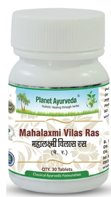
Maha Lakshmivilasa Rasa is a highly revered, classical Ayurvedic mercurial-mineral formulation (Rasa Aushadhi) primarily detailed under the treatment of Kapha disorders (Kapharogachikitsa). This profound formulation balances all three Doshas- specifically pacifying aggravated Kapha and Vata- while deeply kindling the metabolic fire (Agni). It possesses strong immunomodulatory, anti-inflammatory, mucolytic, and tissue-rejuvenating dynamics. Beyond resolving deep-seated respiratory blockages, chronic cough (Kasa), and sinusitis (Pinasa), it penetrates deep tissues to address systemic wasting (Rajayakshma), metabolic errors like diabetes (Prameha), and skin conditions (Kustha) by breaking down advanced cellular blockages (Srotorodha).
CLASSICAL INDICATION
- Kapharoga (Diseases due to Kapha dominance)
- Kasa (Cough)
INGREDIENTS
पलं वज्राभ्रचूर्णस्य तदर्धं गन्धको भवेत् ।
तदर्धं वङ्गभस्मापि तदर्धं पारदस्तथा ॥ 17 ॥
तत्समं हरितालञ्च तदर्धं ताम्रभस्मकम् ।
रसतुल्यञ्च कर्पूरं जातीकोषफलं तथा ॥ 18 ॥
वृद्धदारकबीजस्य बीजं स्वर्णफलं च ।
प्रत्येकं कार्षिकं भागं मृतस्वर्णस्य शाणकम् ॥ 19 ॥
(Rasendrasarasangraha, Kapharogachikitsa; 17–19)
- Vajrabhra churna-bhasma (abhraka) [Bio-purified Mica Calx]
- Gandhaka-shuddha [Purified and Processed Sulfur]
- Vanga-bhasma [Bio-purified Tin Calx]
- Parada-shuddha [Purified and Processed Mercury]
- Haritala-shuddha [Purified Orpiment]
- Tamra-bhasma [Bio-purified Copper Calx]
- Karpura [Cinnamomum camphora extract]
- Jatikosha [Mace – Aril of Myristica fragrans]
- Jatiphala [Nutmeg – Seed of Myristica fragrans]
- Vriddha daraka-bija [Argyreia speciosa seeds]
- Svarna [Bio-purified Gold Calx]
- Mrita svarna bhasma [Calcined Gold Calx]
INGREDIENT DESCRIPTION
Vajrabhra Bhasma (Bio-purified Mica Calx / Biotite Mica)
Serves as a premier cellular rejuvenator (Rasayana) for the respiratory system (Pranavaha Srotas). It exhibits an exceptional ability to penetrate deep tissues (Sukshma), repairing cellular damage caused by chronic infections and wasting conditions like Rajayakshma. Its binding nature stops tissue degeneration and restores cellular immunity. Within this formulation, its primary contribution is providing the core structural defense, ensuring that while other ingredients clear out active disease, the underlying tissue integrity is fortified against long-term deterioration.
Shuddha Parada (Purified and Processed Mercury)
Functions as the essential mercurial core of this formulation. When properly processed, it acts as a supreme catalyst (Yogavahi) that possesses the unique ability to rapidly carry the therapeutic properties of all accompanying herbs and minerals deep into the cellular levels of the body. Its contribution to Maha Lakshmivilasa Rasa is foundational; it enhances the overall bioavailability, accelerates the onset of action, and ensures that the heavy mineral compounds can cross deep biological barriers effectively.
Shuddha Gandhaka (Purified and Processed Sulfur)
Plays a critical role in neutralizing the fierce nature of mercury while amplifying the formulation’s overall therapeutic safety and potency. It acts as a powerful blood purifier and anti-microbial agent that digests systemic metabolic endotoxins (Ama). In this formulation, it works in tandem with Parada to form the Kajjali matrix, providing the necessary anti-inflammatory and detoxifying backbone required to combat stubborn Kapha infections.
Vanga Bhasma (Bio-purified Tin Calx)
Systematically targets Kapha accumulations and is particularly renowned for strengthening the genitourinary tract and addressing conditions like Prameha (diabetes/metabolic errors). Its contribution to the formulation is to stabilize fluid metabolism and prevent the systemic depletion of vital essences (Ojas). By regulating moisture and tissue metabolic rates, it ensures that the body does not yield to the heavy, stagnant dampness characteristic of chronic Kapha disorders.
Shuddha Haritala (Purified Orpiment)
Offers strong anti-microbial and anti-pyretic action, clearing deep-seated toxins within the blood and skin tissues, which aids in treating severe skin diseases (Kustha) and chronic sinuses (Nadivrana). Its inclusion significantly upgrades the formulation’s capability to fight deep-rooted, chronic infections and infectious pathways. It helps break down stubborn biofilms and deep-seated cellular impurities that standard herbal mixtures cannot reach.
Tamra Bhasma (Bio-purified Copper Calx)
Acts as a potent scraping agent (Lekhana), dissolving thick, sticky phlegm in the lungs and reducing adipose tissues in Sthaulya (obesity). Its specific contribution to this formulation is its powerful direct action on congested channels; it actively carves through and eliminates dense, stagnant mucus and lipid overloads, effectively opening up the respiratory and circulatory passages for free-flowing vitality.
Karpura (Camphor – Cinnamomum camphora extract)
Provides volatile aromatic properties that act as instant expanders of the respiratory channels. It relieves throat irritation (Galaroga), clears sinus congestion, and possesses soothing, anti-spasmodic properties that control pain (Shula). In Maha Lakshmivilasa Rasa, Karpura contributes immediate, cooling relief to inflamed airways and acts as a localized antiseptic, quickly easing bronchospasms and refreshing the upper respiratory tract.
Jatikosha (Mace – Aril of Myristica fragrans)
Delivers sharp, aromatic, and heating dynamics that immediately help clear throat irritation (Galaroga), neutralize mouth odor (Mukharoga), and dispel accumulated head-congestion. Its role in the formulation is to optimize upper respiratory ventilation and enhance digestive fire, preventing the stomach from generating further Ama that could rise and exacerbate lung congestion.
Jatiphala (Seed of Myristica fragrans)
Brings a highly specialized combining action that grounds the formulation, soothing the nervous system while providing anti-spasmodic relief to the respiratory muscles. It works synergistically with the volatile oils of the other spices to control pain (Shula) and halt chronic, hacking coughs. Its presence ensures that the formulation calms hyperactive airways while drying up excessive, runny secretions.
Vriddhadaru (Argyreia speciosa seeds)
Is an exceptional tissue vitalizer and nervous system tonic. Its contribution to this compound is to counteract the physical exhaustion and neurological stress brought on by chronic coughing and systemic illness. By strengthening the nervous system and supporting cellular regeneration, it ensures the patient regains stamina and physical resilience during recovery.
Svarna Bhasma (Bio-purified Gold Calx)
Is an elite tissue vitalizer that directly improves cardio-respiratory endurance, acts as a powerful aphrodisiac (Vajikarana), balances all three Doshas, and protects vital organs from the stress of chronic inflammation and structural wasting. It elevates Maha Lakshmivilasa Rasa into a premium, top-tier remedy by acting as a shield for the heart and lungs, boosting natural immunity (Vyadhikshamatva), and restoring a radiant life-force to a depleted physiology.
Mrita Svarna Bhasma (Calcined Gold Calx)
Acts as an intensely refined, highly potent form of gold calx that further amplifies the formulation’s life-saving, cellular-rejuvenating dynamics. It specifically targets the deep immune response and prevents cellular collapse in advanced, wasting diseases. Its elite contribution is to step in as an ultimate bio-protective agent, accelerating tissue repair, optimizing cardiac tone, and reversing deep degenerative damage at a rapid pace.
METHOD OF PREPARATION
To prepare Maha Lakshmivilasa Rasa, first combine purified Mercury (Shuddha Parada) and purified Sulfur (Shuddha Gandhaka) to create a fine, lusterless black powder (Kajjali). Gradually introduce the remaining mineral components- Vajrabhra Bhasma, Vanga Bhasma, Tamra Bhasma, and Shuddha Haritala- into the mortar, incorporating them through sustained mechanical trituration.Separately pulverize the plant-based materials- Karpura, Jatikosha, Jatiphala, and Vriddhadaru- into an ultra-fine blend and mix them with the core mineral compound alongside Svarna Bhasma. Wet-triturate the mixture thoroughly with designated liquid mediums until it achieves a smooth, unified, dough-like consistency.
निष्पिष्य वटिका कार्या द्विगुञ्जाफलमानतः ।
(Rasendrasarasangraha, Kapharogachikitsa; 19 ½)p
This text directs the precise processing and therapeutic density of the medicine, stating: “Upon thorough grinding and compounding, uniform pills must be rolled equivalent to the weight of two Gunja seeds (250 mg).”
Roll the mass into uniform 250 mg pills (Dvigunja size), dry them under clean shade, and preserve them inside airtight glass containers.
MEDICINAL PROPERTIES
For Kasa And Pinasa (Chronic Cough, Bronchitis, Sinusitis, and Rhinitis)
Administer Maha Lakshmivilasa Rasa along with Sitopaladi Churna or Talisadi Churna. The best adjuvants for this condition are honey or lukewarm water. This targeted herbo-mineral formulation uses its sharp, deep-penetrating, and volatile aromatic properties to clear stubborn upper respiratory blocks, liquefy dense phlegm, soothe inflamed bronchial passages, and dry up chronic nasal secretions to restore clear airways.
For Rajayakshma (Tuberculosis and Chronic Pulmonary Wasting)
Administer Maha Lakshmivilasa Rasa along with Chyawanprash or Ashwagandha Churna. The best adjuvant for this condition is warm milk. This premium formulation utilizes its elite cell-rejuvenating (Rasayana) and tissue-building attributes to arrest rapid tissue decay, rebuild compromised muscular strength, improve cardio-respiratory endurance, and stimulate cellular immunity to counter deep-seated system exhaustion.
For Sthaulya And Amavata (Obesity, Metabolic Congestion, and Rheumatoid Arthritis)
Administer Maha Lakshmivilasa Rasa along with Amritadi Guggulu or Simhanada Guggulu. The best adjuvants for this condition are warm water or honey. This potent combination utilizes deep-scraping (Lekhana) mineral calces to dismantle stubborn metabolic endotoxins (Ama), digest accumulated fat deposits, reduce painful structural joint swelling, and clear cellular channels to relieve chronic musculoskeletal stiffness.
For Vajikarana (Diminished Reproductive Vitality and Sexual Debility)
Administer Maha Lakshmivilasa Rasa along with Kapikachhu Churna or Musli Pak. The best adjuvant for this condition is warm milk. This mineral-dense formula infuses pure Svarna Bhasma to nourish the deepest reproductive tissues (Shukra Dhatu), elevate nervous system stamina, restore lost vitality, and enhance absolute vigor across the entire physiology.
For Galaroga And Mukharoga (Throat Diseases, Tonsillitis, and Oral Mucosal Disorders)
Administer Maha Lakshmivilasa Rasa along with Khadiradi Vati or allow the pill to dissolve slowly in the mouth. The best adjuvants for this condition are a decoction of Yashtimadhu (licorice) or honey. This combination releases direct antimicrobial and anti-inflammatory properties into the oral cavity to immediately neutralize localized pathogens, soothe burning throat irritation, reduce vocal cord edema, and clear toxic coatings from the tongue.
For Kustha And Tvakroga (Stubborn Skin Disorders, Eczema, and Psoriasis)
Administer Maha Lakshmivilasa Rasa along with Kaishore Guggulu or Mahamanjisthadi Kwatha. The best adjuvant for this condition is lukewarm water. This powerful blood-purifying formulation utilizes its arsenic-purified and mercurial compounds to penetrate deep plasma-tissue beds, eradicate circulating toxins, neutralize systemic heat, and halt chronic epidermal scaling and stubborn itching.
For Nadivrana And Bhagandara (Fistulous Tracts, Deep Sinuses, and Fistula-in-Ano)
Administer Maha Lakshmivilasa Rasa along with Triphala Guggulu or Kanchanar Guggulu. The best adjuvants for this condition are warm water or a decoction of Khadiara. This formulation drives intense anti-microbial and tissue-healing actions to scrape out necrotic tissue from deep tracts, reduce chronic purulent discharge, stimulate healthy granulation, and close complex internal wounds from within.
For Shula (Colic, Neuralgic Spasms, and Acute Internal Pain)
Administer Maha Lakshmivilasa Rasa along with Sankha Vati or Maha Shankha Vati. The best adjuvants for this condition are warm water or honey. This formulation combines volatile essential oils and grounding minerals to regulate hyperactive nervous impulses, break up trapped gas pockets, release visceral smooth muscle spasms, and calm intense, radiating internal pain.
IMPACT ON DOSHA
Maha Lakshmivilasa Rasa exhibits a strong Kapha-Vata pacifying action. Its scraping (Lekhana), channel-clearing (Srotoshodhana), and heating dynamics liquefy and dry up excess Kapha, while its mineral bases ground and balance unstable Vata.
INDICATIONS
- Kasa (Cough) & Pinasa (Sinusitis/Rhinitis)
- Rajayakshma (Tuberculosis)
- Sthaulya (Obesity) & Amavata (Rheumatoid Arthritis)
- Vajikarana (Diminished Reproductive Vitality)
- Galaroga (Throat diseases) & Mukharoga (Mouth diseases)
- Kustha & Tvakroga (Stubborn Skin Disorders)
- Nadivrana (Fistulous tracts) & Bhagandara (Fistula-in-ano)
- Shula (Colic)
DOSAGE
250 mg (equivalent to 2 Gunja pills), taken once or twice a day as directed by an Ayurvedic physician.
ANUPANA (Adjuvant)
Honey or Milk.
CONCLUSION
Maha Lakshmivilasa Rasa is an extraordinary herbo-mineral masterpiece that showcases the depth of Ayurvedic alchemy (Rasashastra). By targeting the root of Kapha-Vata stagnation, it effectively clears respiratory pathways, eliminates systemic toxins, and halts chronic degenerative processes. Its dual capacity to scrape away deep metabolic waste while simultaneously providing cell-rejuvenating Rasayana benefits makes it an invaluable clinical remedy for complex, multi-system disorders.